News
John Desmond Bernal
Posted by: Joachim Frank |
January 11, 2017 |
No comment
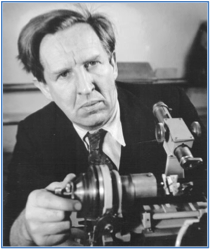 In Tallahassee, Florida, I had the privilege to participate in the 90th birthday celebration for Don Caspar, as I will report in another note. Andrew Brown, the last speaker of that Symposium organized at Florida State University gave a historical account of J D Bernal, a physicist who crossed paths with Don early on and must have been quite influential for his thinking.
Now what interested me greatly is that Bernal, as early as 1931, well before the discovery...
In Tallahassee, Florida, I had the privilege to participate in the 90th birthday celebration for Don Caspar, as I will report in another note. Andrew Brown, the last speaker of that Symposium organized at Florida State University gave a historical account of J D Bernal, a physicist who crossed paths with Don early on and must have been quite influential for his thinking.
Now what interested me greatly is that Bernal, as early as 1931, well before the discovery...Read More
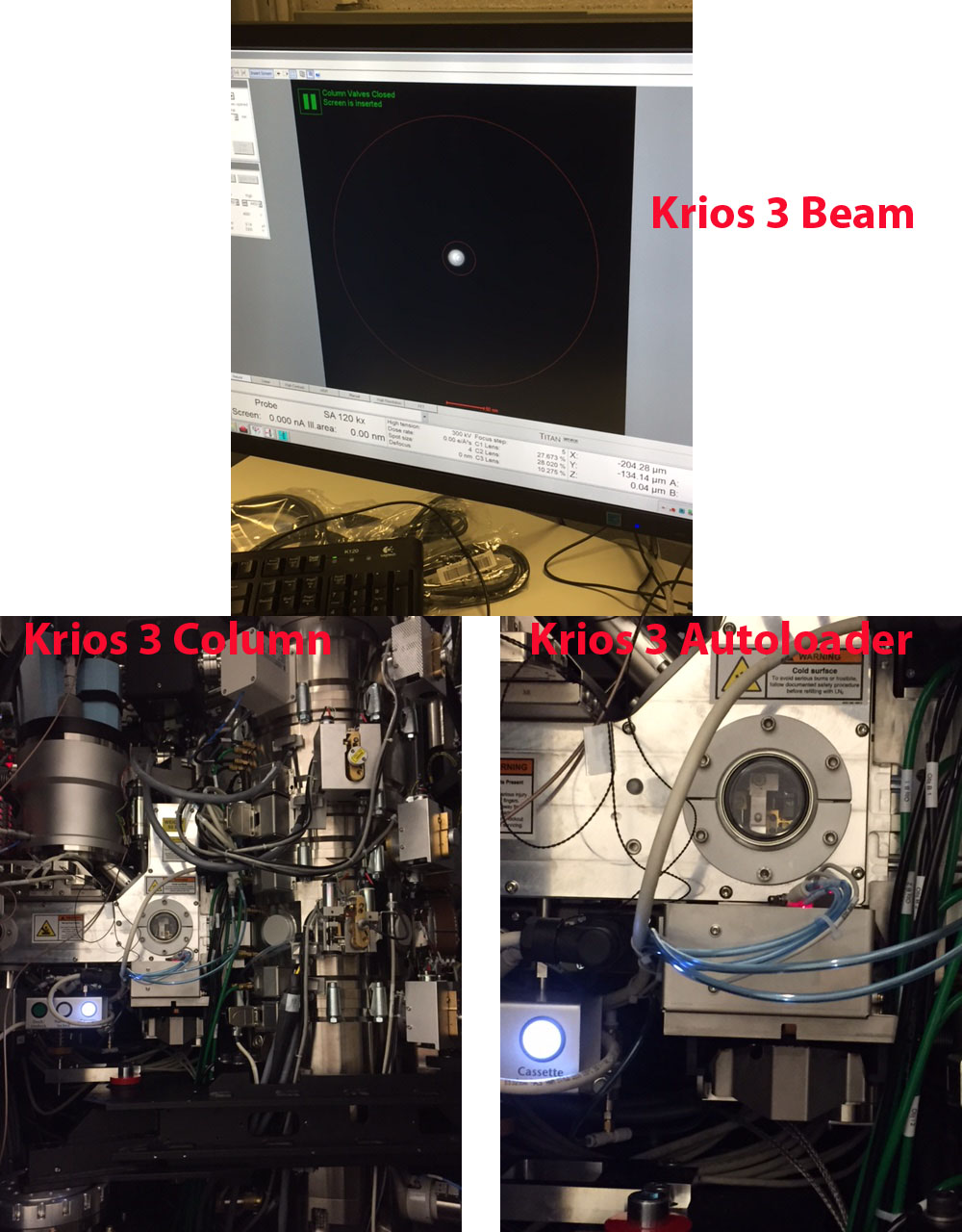
The Beam is On!
Posted by: Joachim Frank |
January 5, 2017 |
No comment
Read More
New article in Nature Protocols
Posted by: Joachim Frank |
January 5, 2017 |
No comment
Read More
Commentary to Structure, Fu et al.
Posted by: Joachim Frank |
January 2, 2017 |
No comment
Read More
Klaus Schulten
Posted by: Joachim Frank |
November 3, 2016 |
No comment
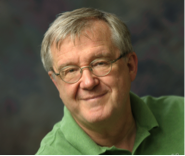 I was very sad to hear that Klaus Schulten passed away on November 1 after several months of illness. He was an enthusiastic collaborator and friend for more than a decade. We had just submitted a joint paper on a general method of describing domain motions in molecular machines for the Festschrift planned for the celebration of his 70th birthday. Our earlier collaborations, in which his students Elizabeth Villa and Leonardo Trabuco played...
Read More
New paper in PNAS
Posted by: Joachim Frank |
October 11, 2016 |
No comment
 Our paper describing the 2.5-Å structure of the large ribosomal subunit of T. cruzi has finally appeared.
It presents a culmination of a collaboration of four groups: our own lab, Susan Madison Antennucci’s at the Wadsworth Center in Albany, Liang Tong’s at the Department of Biological Sciences, also Columbia University, and John Woolford’s at Carnegie Mellon University in Pittsburgh.
“Structure and assembly...
Our paper describing the 2.5-Å structure of the large ribosomal subunit of T. cruzi has finally appeared.
It presents a culmination of a collaboration of four groups: our own lab, Susan Madison Antennucci’s at the Wadsworth Center in Albany, Liang Tong’s at the Department of Biological Sciences, also Columbia University, and John Woolford’s at Carnegie Mellon University in Pittsburgh.
“Structure and assembly...Read More
Structural Basis for Gating and Activation of RyR1
Posted by: Joachim Frank |
September 23, 2016 |
No comment
Read More
Congratulations Dr. Ming Sun
Posted by: Joachim Frank |
July 19, 2016 |
No comment
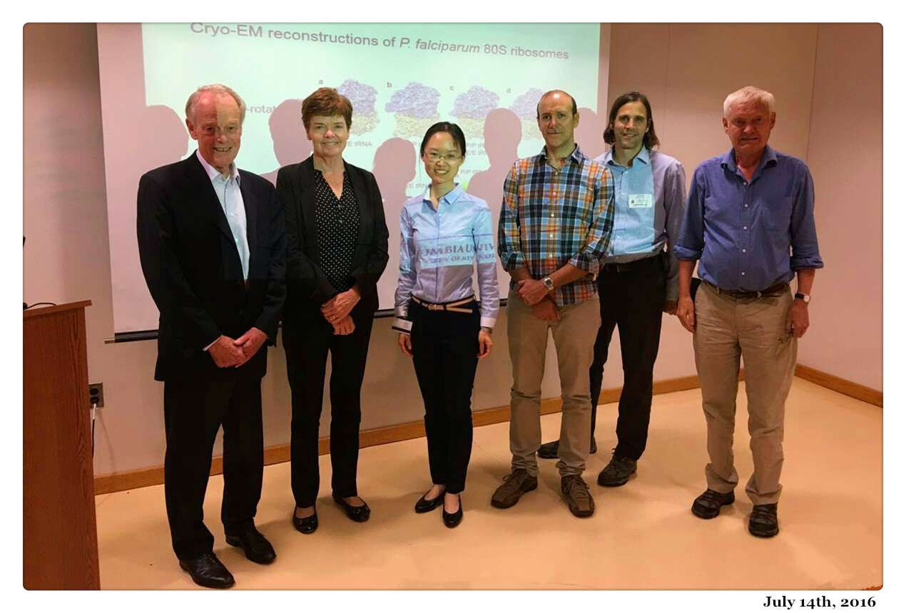 Congratulations to Dr. Ming Sun for her successful thesis defense on Thursday, July 14, 2016.
The title of her Ph.D. thesis is “Cryo-Electron Microscopy Studies of Dynamical Features of Ribosomes During the Translation Process.”
Members of her Thesis Committee: Ruben Gonzalez (Chair), Joachim Frank (Sponsor/Mentor), Wayne Hendrickson, Bridget Carragher, and Jeffrey Dvorin (Children’s Hospital, Harvard Medical School)
Congratulations to Dr. Ming Sun for her successful thesis defense on Thursday, July 14, 2016.
The title of her Ph.D. thesis is “Cryo-Electron Microscopy Studies of Dynamical Features of Ribosomes During the Translation Process.”
Members of her Thesis Committee: Ruben Gonzalez (Chair), Joachim Frank (Sponsor/Mentor), Wayne Hendrickson, Bridget Carragher, and Jeffrey Dvorin (Children’s Hospital, Harvard Medical School)Read More
New paper accepted by Cell
Posted by: Joachim Frank |
July 1, 2016 |
No comment
Read More
New paper in Science
Posted by: Joachim Frank |
July 1, 2016 |
No comment
Alexander I. Sobolevsky. Abstract: AMPA-subtype ionotropic glutamate receptors (AMPARs) mediate fast excitatory neurotransmission and contribute to high cognitive processes such as learning and memory. In the brain, AMPAR trafficking, gating, and pharmacology is tightly controlled by transmembrane AMPAR regulatory proteins...
Read More


