News
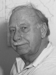
Donald Parsons
Posted by: Joachim Frank |
December 31, 2017 |
No comment
 Donald Parsons passed away earlier this month at age 89. He established high-voltage electron microscopy at the Division of Labs and Research of the New York State Department of Health in Albany, later to be renamed Wadsworth Center.
Parsons was convinced that most cancers were caused by viruses, and that the reason they had not been found was a sampling problem. Imaging whole cells in 3D with 1MV electrons would be a way to find them.
Parsons hired me in 1975 to establish an image processing group. The high-voltage microscope was still in crates when I arrived, and together with William Goldfarb (whom I hired, once in Albany) and a couple of students I started out with the development of an image processing suite, SPIDER, to implement single-particle techniques as well as electron tomography.
He was an early visionary, much ahead of his time in thinking about the construction of a navigable high-resolution model of a whole cell based on tomographic reconstructions. Only now, 40 years later, we have the technical capabilities to realize this idea.
Another pioneering move was the construction of an environmental chamber, which allowed fully hydrated samples to be imaged in the high-voltage electron microscope. Although little came out of it because of technical problems, his early contribution to the solution of the basic incompatibility problem posed by biological matter in vacuum should be duly acknowledged.


