News
Miloslav (“Milas”) Boublik 1927 – 1994
Posted by: Joachim Frank |
January 16, 2019 |
No comment

In my recent talk at the Ribosome meeting in Merida, Mexico, which traced the role of the ribosome in driving and facilitating the development of single-particle EM, I paid tribute to Milas Boublik. At the time I met him, in 1980, he was a research scientist at Roche, in Nutley, New Jersey. This institute of Roche has since been closed.
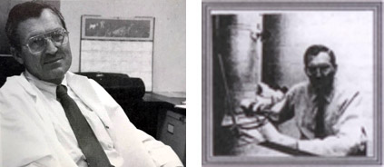
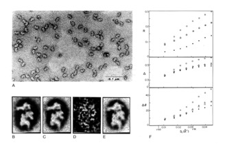
The image on the left was sent to me by Martin Kessel, who contacted Herb Weissbach, one of the former Directors of the Institute. The grainy one on the right is from a flyer published by the Structural Biology Group at the Wadsworth Center. It appeared in an obituary written by Adriana Verschoor, who was part of my group. Again, I have to thank Martin for retrieving the scan of the flyer from his archive.
After a lecture I gave at the Rockefeller University describing my ideas about single-particle techniques, Milas introduced himself to me and showed me his micrographs of negatively stained HeLa ribosomes. Would they be of interest in applying my method? I seized this opportunity because the particles showed two preferential views (the “L” and “R” views), convenient for developing and demonstrating image alignment and two-dimensional averaging. This was my first introduction to the ribosome. It led to the publication of my first paper in Science in 1981. From that time on, until the present day, the quest for the structure of the ribosome was a strong motivating force in the development of single-particle reconstruction.


